梅斯医学MedSci APP
医路相伴,成就大医


梅斯医学MedSci APP
医路相伴,成就大医
早期复极心电图改变及J波常见于特发性室颤的患者,其发生率明显高于正常人群。近来的临床研究表明,早期复极心电图改变是特发性室颤患者发生室颤及心脏性猝死的预测因素,还有研究表明早期复极合并ST段水平型或压低与恶性心律失常发生强烈相关,因此早期复极心电图改变是特发性室颤患者的特征性心电图改变。
然而仍有许多特发性室颤患者并没有早期复极心电图改变,是否还有其他心电图特征目前还不清楚,还需要进一步临床研究。据此,日本学者Sekiguchi Y等进行了一项临床研究,近来发表于JCE杂志,该研究旨在探讨特发性室颤新的心电图特征。
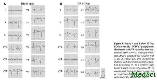
该研究共入选64例特发性室颤患者,所有患者均来自日本特发性室颤注册研究,所有患者至少经历一次室颤发作,且排除了器质性心脏病及Brugada综合征,根据患者是否存在早复极心电图改变分成两组,早复极心电图改变定义为下壁或侧壁J点抬高大于0.1mv。其中存在早复极心电图改变的患者有24例,占38%;无早复极心电图改变的患者有40例,占62%。存在早复极心电图改变的患者绝大多数为男性,占92%,而无早复极心电图改变的患者男女基本均等。存在早复极改变的患者心电图均无室内传导异常、电轴异常或束支传导阻滞。而无早复极改变的患者中有9例存在室内传导障碍,且这些患者的PR间期及QRS波时限均长于无室内传导障碍的患者。
根据该项研究可得出以下结论:目前发现特发性室颤的患者存在3种心电图特征:早复极心电图改变、室内传导障碍、PR间期延长,对于不存在早复极心电图改变的患者,室内传导障碍是特发性室颤新的临床特征。
New Clinical and Electrocardiographic Classification in Patients with Idiopathic Ventricular Fibrillation.
INTRODUCTION
The presence of early repolarization (ER) recently has been considered as a prognostic marker for sudden cardiac death in patients with idiopathic ventricular fibrillation (IVF), but there are certain numbers of IVF patients lacking ER. We aimed to clarify the clinical and electrocardiographic characteristics of the patients with IVF in the presence and absence of ER.
METHODS AND RESULTS
We studied 64 consecutive IVF patients from the Japan Idiopathic Ventricular Fibrillation Study (J-IVFS) registry, which subjected with at least one episode of documented VF in the absence of structural heart diseases and excluding Brugada syndrome. We assessed clinical and electrophysiological characteristics in the IVF patients with and without ER. ER was defined as J-point elevation of >0.1 mV in either inferior or lateral leads. Twelve-lead electrocardiogram (ECG) demonstrated 24 (38%) of 64 patients with ER (ER[+] group) and the remaining 40 (62%) patients without ER (ER[-] group ). ER[+] group had a male predominance (92% for males) and ER[-] group revealed nearly equal distribution in both sexes. While no patients in ER[+] group showed intraventricular conduction disturbance (CD) with abnormal axis deviation and/or bundle branch block in ECG, 9 in ER[-] group had signs of CD (ER[-]/CD[+] subgroup). ER[-]/CD[+] subgroup had prolonged P-R interval and QRS duration compared to other patient groups.
CONCLUSION
We found 3 distinct ECG patterns in IVF patients. In addition to the presence and absence of ER, there is a subgroup without ER demonstrating intraventricular CD, which represents a distinct clinical entity of IVF.